

Dear Sir,
The flexor retinaculum (FR), also known as the transverse carpal ligament, is a thick fibrous band that forms the superficial border of the carpal tunnel.(1-3) It has medial (ulnar) attachments to the hook of the hamate (HoH) and pisiform and lateral (radial) attachments to the trapezium tubercle and scaphoid.(1-3) HoH injuries are uncommon; to our knowledge, isolated injury of the FR at its attachment to the HoH has not been previously described. We herein present the first case report of an isolated tear of the FR at its HoH attachment, which was diagnosed on magnetic resonance (MR) imaging.
A 27-year-old man presented with pain in the left wrist after a fall. The patient had tripped and fallen forward, sustaining a contusion on his outstretched hand. On physical examination, there was localised tenderness at the HoH. Radiographs of the wrist with dedicated view of the carpal tunnel showed an intact HoH with no fracture or dislocation (
Fig. 1
Radiograph of the wrist in carpal tunnel view shows no abnormality at the hook of the hamate.
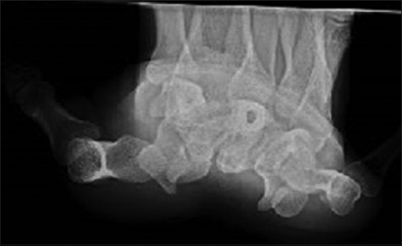
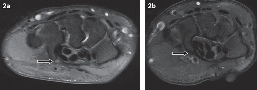
Fig. 2
(a) Axial proton-density fat-saturated (PD FS) MR image of the left wrist shows linear increased signal of the flexor retinaculum at its attachment to the hook of the hamate, compatible with a partial tear (arrow). (b) Axial PD FS MR image of another patient shows no symptoms localised to the hook of the hamate. The normal, uniformly low signal of the attachment of the flexor retinaculum to the hook of the hamate can be appreciated (arrow). Both images were obtained with the same 1.5 T scanner.
The FR is identified on MR imaging as a uniformly hypointense structure on all sequences (due to its fibrous composition) and is thickest at its medial attachments.(1-3) It performs the primary function of enclosing the flexor tendons in the carpal tunnel.(2) Pathology of the FR is most commonly related to carpal tunnel syndrome, in which palmar bowing of the FR can be seen as a secondary imaging feature in addition to hyperintense swelling of the median nerve.(4) There is limited literature on the imaging appearance of acute injury to the FR.
The HoH is an uncommon site of pathology in the wrist, with fractures being the most commonly encountered type.(3,5) Other entities that can affect the HoH include bipartite hamate, pisiform-hamate coalition, infection, avascular necrosis and tumours.(3,6) Fractures of the HoH are often missed on both clinical examination and imaging, as clinical presentation can be non-specific,(3) especially in the presence of concomitant injuries such as a scaphoid fracture.(7) In the setting of a suspected radiographically occult HoH fracture, computed tomography (CT) is often performed.(3) MR imaging is preferred for the evaluation of concomitant soft tissue injuries, such as those of the flexor tendons or ulnar nerve.(3)
Prior to this case, we had not observed similar MR findings of isolated linear hyperintense signal at the FR attachment to the HoH, including in scans of other patients performed on the same machine with the same scan protocol. There was consistently uniform low signal of the FR at its attachment to the HoH. An extensive literature review also did not identify any previously reported cases. We believe that this novel MR finding represents a focal tear or sprain. The finding was important in our case because it prevented the patient from undergoing a potential surgical intervention.
Our case demonstrates that in addition to a HoH fracture, isolated tears of the FR attachment can also cause significant pain and disability. This finding should thus be actively sought in patients who undergo MR imaging after injuries to the wrist, especially if there are symptoms localised to the HoH. However, this can be a subtle finding if it is an isolated injury, in the absence of accompanying soft tissue or marrow oedema. Indeed, in our patient, the partial tear was visualised only on a single axial slice and could easily have been overlooked. This should thus be a review area, with the reviewing radiologist searching for any linear hyperintense signal emanating from the FR attachment to the HoH on fluid-sensitive sequences. We term this phenomenon the ‘hook line’ sign or ‘Salil-Zhang-Abhinav’ sign.
In our case, the mechanism of injury was blunt trauma after a fall on an outstretched hand, which is a common cause of HoH fractures.(3) We believe that other mechanisms that predispose a person to HoH fractures, such as repetitive microtrauma or avulsion injury, can also lead to isolated injury to the FR attachment. We postulate that this injury was not previously identified on imaging because CT (instead of MR imaging) is often used for evaluation after initial radiographs are negative for HoH fracture. MR imaging has the advantage of improved diagnosis of soft tissue injuries, including isolated FR tear, notwithstanding its increased cost and less widespread availability. Conservative treatment should suffice in cases of isolated FR tear, such as in our patient, who had good functional recovery.
In summary, we present the first reported case of isolated tear of the FR at its attachment to the HoH, which was diagnosed due to the ‘hook line’ sign, a novel MR finding. In the absence of HoH fracture, such a tear may be the cause of persistent pain and disability after trauma and should be actively sought on MR imaging.
Yours sincerely,