

Dear Sir,
We herein report a case of a 63-year-old woman who presented with shortness of breath and palpitations. Electrocardiography showed fast atrial fibrillation and blood tests revealed iron-deficiency anaemia. Optical colonoscopy revealed an ascending colon tumour. Chest radiography showed retrocardiac atelectasis in the left lower lobe (
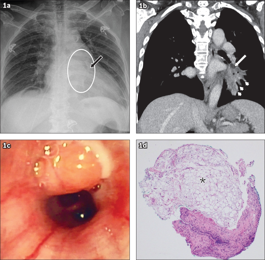
Fig. 1
(a) Frontal chest radiograph shows a vague opacity in the left lower zone (black arrow) with a smooth margin, which appears to protrude into the bronchial lumen. There is retrocardiac opacification, suggestive of post-obstructive atelectasis (circled). (b) Coronal mediastinal window multidetector computed tomography image shows that the nodule has fat attenuation (white arrow) and no calcification. Post-obstructive atelectasis is present (arrowheads). (c) Bronchoscopic image shows a lobulated fleshy lesion at the orifice of the left lower lobe basal segments, occluding the anterobasal segment of the left lower lobe. Endobronchial biopsy caused extrusion of milky contents from the lesion. (d) Photomicrograph shows bronchial tissue covered by benign epithelium, benign seromucinous glands and mature adipose tissue (asterisk) (Haematoxylin & eosin, × 5).
Endobronchial lipomas account for 0.1%–0.5% of lung tumours(1) and about 13% of benign lung tumours.(2) Patients present in middle age and are usually male, with a strong male predominance (male-to-female ratio of 45:7).(3) Clinically, these patients most often present with cough and symptoms of chest infection.(2) Their diagnosis is often delayed due to the indolent nature of this tumour. Some investigators have found that smoking and obesity are correlated as risk factors for endobronchial lipoma.(2,4) However, although our patient was overweight, she was a non-smoker.
MDCT with isotropic resolution and thin slice thickness (ideally 1 mm) is the imaging modality of choice. Chest radiography is less useful, as it has low sensitivity of about 65%.(5) MDCT defines intraluminal and extraluminal extent, as well as potential complications such as obstructive atelectasis. Assessment of tumour density is also important. The presence of fat attenuation narrows down the differential diagnoses to the following: pure endobronchial lipoma, which demonstrates homogeneous fat attenuation; fibrolipomatous tumour, which contains soft tissue attenuation with islands of fat; and hamartoma, which has fat density alternating with calcific foci.
Endobronchial lipoma may be managed by endoscopic resection or surgical excision. Endoscopic resection is usually the first line of treatment, as it is both diagnostic and curative. Furthermore, endoscopic resection is less invasive, conferring a lower morbidity rate than surgical resection, and effectively relieves symptoms.(4) However, many of these tumours grow through the bronchial wall, making complete endoscopic resection difficult. Recurrence of endobronchial lipoma is exceedingly rare after complete surgical resection. Following resection, the need for endoscopic surveillance is debatable. MDCT of the thorax with virtual bronchoscopy provides a noninvasive approach, but is limited in that the distal airways may prove difficult to visualise.
In conclusion, endobronchial lipoma is a rare benign lung tumour that presents late due to nonspecific symptoms. A multidisciplinary approach is crucial for successful management.
Yours sincerely,
ACKNOWLEDGEMENTS
The authors thank Dr Lwin Khin Yu and Dr Michael Tan from the Department of Pathology, Changi General Hospital, Singapore, for their help in obtaining histopathological slides for this publication.