

Dear Sir,
Clinical bedside evaluation of swallowing is the first procedural step that a clinician who is treating dysphagia uses to evaluate the swallowing function of a patient. This involves using the bare hands to palpate the neck to identify key landmarks such as the thyroid, hyoid, cricoid and the base of the tongue. The hyoid bone is a U-shaped structure situated at the anterior part of the neck between the jaw bone and the thyroid cartilage, and it is secured to the thyroid by ligaments. The hyoid bone also serves as the point of attachment for several muscles that are involved in the swallow function.
As part of the professional curriculum, clinicians learn the four-finger placement(1) for palpation of the neck while assessing a patient’s swallow. Some clinicians are also taught to identify the hyoid by locating the greater and lesser horns of the hyoid.
Based on our experience conducting training programmes across the region over the years, we recognise that clinicians often tend to overlook the location of the hyoid or misidentify it. This has consequences and implications for not only the assessment findings, but also the eventual treatment plan, which may involve procedures that require working with muscles in the suprahyoid and infrahyoid regions during dysphagia management.
The S-Hyoid Finder is a simple, novel method proposed by the main author and validated by both the authors with normal individuals. The process involves the following steps:
-
Have the patient sit in an upright or reclined position, with the head and neck supported upright on the bed.
-
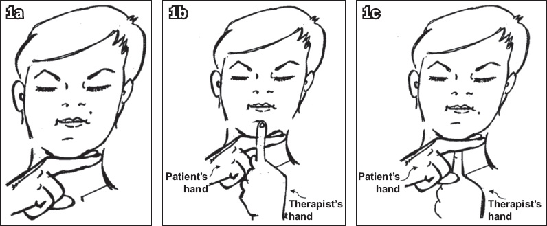
Place the patient’s forefinger and middle finger together under the chin exactly at the margin of the mandible (
Fig. 1a). -
The clinician then runs down his/her forefinger along the midline of the patient’s face, over the mandible and patient’s fingers that are placed under the mandibular margin (
Fig. 1b). -
The clinician’s forefinger then lands on a small depression/supple area immediately posterior to the patient’s fingers (
Fig. 1c). -
The clinician then palpates gently in that depression and feels for the hyoid in that space.
-
By asking the patient to swallow saliva, the clinician can then sense the hyoid movement upward, forward and downward post swallow.
-
Keeping the middle finger on the identified spot, the clinician moves the forefinger above to palpate the base of the tongue, the ring finger below on the thyroid notch and the little finger on the cricoid (four-finger placement), and clinically assesses the swallow again.
Fig. 1
Illustrations show the process of identifying the hyoid bone using the S-Hyoid Finder method.
As a way of validating the accuracy of this method, the dysphagia clinicians on the team cross-checked to evaluate if the position was the same as that identified by the traditional palpation and identification methods. A 100% congruence between the two techniques was noted while examining normal and dysphagic patients in both clinical practice and training sessions. We propose that a radiological study could be conducted to further evaluate the S-Hyoid Finder method, as it would serve as an easy and precise tool for both novice and experienced clinicians.
Yours sincerely,