

Abstract
The palmaris longus is harvested as a tendon graft in various surgical procedures. We herein report the variations in the insertion of the palmaris longus tendon. During a routine dissection, a rare variation in the insertion of the palmaris longus tendon was observed. In the left forearm, the palmaris longus tendon bifurcated, while in the right forearm, the palmaris longus tendon trifurcated, giving rise to an accessory muscle, which passed superficial to the ulnar artery and ulnar nerve. The accessory muscle was supplied by a deep branch of the ulnar nerve, and the ulnar artery was observed to be tortuous. During reconstructive surgeries, surgeons should bear in mind the accessory muscle. Also, since the palmaris longus muscle provides a very useful graft in tendon surgery, every surgeon should be aware of the variations in the insertion of the palmaris longus tendon.
INTRODUCTION
The palmaris longus is often described as one of the most variable muscles in the human body, and is phylogenetically classified as a retrogressive muscle (i.e. muscle with a short belly and a long tendon).(1) The palmaris longus is a slender fusiform muscle medial to the flexor carpi radialis muscle, and arises from the medial epicondyle by the common flexor tendon, the adjacent intermuscular septa and the antibrachial fascia. Its long, slender tendon passes in front of the flexor retinaculum and is continuous with the central part of the palmar aponeurosis.(2)
Tendon grafts are frequently needed in reconstructive surgery of the hand. Most surgeons agree that the palmaris longus tendon is the first choice as a tendon donor because it fulfils the necessary requirement in length, diameter and availability, and can be used without resulting in any functional deformity. The palmaris longus tendon is often considered the ideal donor of tendon grafts for replacement of long flexors of the fingers and thumb. The superficial position of the palmaris longus and its vestigial nature are other important positive points for its choice as a tendon donor.(3)
In vertebrates, the palmaris longus is found only in mammals and is best developed in those where the forelimb is used for ambulation. The palmaris longus is variably absent in higher apes such as chimpanzees and gorillas, but is always present in the orangutan. In humans, the absence of the palmaris longus appears to be hereditary, but its genetic transmission is not clear.(3) Herein, we report an interesting case of variation in the insertion of the palmaris longus tendon.
CASE REPORT
During a routine dissection of an adult male cadaver aged about 50 years at the Department of Anatomy, Kempegowda Institute of Medical Sciences, Bangalore, India, a rare variation in the insertion of the palmaris longus tendon in both upper limbs was observed. The region of interest was finely dissected and photographed. In the left forearm, the palmaris longus tendon, which measured 15 cm in length and 0.8 cm in its maximum width, bifurcated toward the insertion (
Fig. 1
Photograph of the left forearm shows the bifurcation of the palmaris longus tendon and the accessory muscle with its nerve supply.
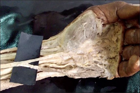
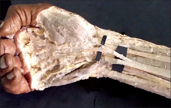
Fig. 2
Photograph of the right forearm shows the trifurcation of the palmaris longus tendon and the accessory muscle with its nerve supply.
DISCUSSION
The palmaris longus may be attached to any of the following structures: the fascia of the forearm, tendon of the flexor carpi ulnaris, flexor retinaculum (superficial or deep), pisiform, scaphoid, the middle phalanx of the fourth digit, short abductor of the thumb, fascia and muscles of the hypothenar eminence, one of the flexor tendons, or in the vicinity of a metacarpophalangeal joint.(4)
The palmaris longus may enter Guyon’s canal and cause compression of the ulnar artery and ulnar nerve located in the canal.(4) The tendon of insertion may split into two or more bundles, in which case the accessory tendon(s) may be inserted into one of the aforementioned structures.(4) Palmaris longus variations have been classified as: (a) complete agenesis; (b) variation in location and form of its fleshy part; (c) aberrancy of attachment at its origin or insertion; (d) duplication and triplication; (e) accessory slips; and (f) replacing elements of similar form or position.(4) The variation of the palmaris longus highlighted in our case report is the aberrancy of attachment of the palmaris longus at its insertion, with the presence of an accessory muscle.
Variations of the palmaris longus muscle also include uni- or bilateral absence in 10% of patients, double palmaris longus muscle with a tendinous cross slip, and bifid reversed palmaris longus muscle. Mobin reported on the use of magnetic resonance imaging to show bilateral accessory palmaris longus muscles, duplication of palmaris longus muscle, anomalous conjoined tendons of palmaris longus muscle, and flexor carpi ulnaris muscle.(5) A number of variations in the origin, course and insertion of the palmaris longus have been noted, including anomalous insertion deep into the retinaculum that causes a carpal tunnel-like syndrome; three-headed reversed palmaris longus causing compartment syndrome; and double palmaris longus.(6) A variant pattern of the insertion of the palmaris longus with the median nerve located medially to the palmaris longus at the wrist has also been demonstrated.(6)
The muscle belly or its tendon, or both, may be bifid. When a cleft is present in the muscle, the tendons are likely to insert separately into the palmar aponeurosis.(7) Although the tendon of the palmaris longus muscle is continuous with the palmar aponeurosis, histological and developmental studies revealed that these two structures arise independently and do not share a common origin. This may explain the variation between the two entities with regard to attachment areas.(8)
The palmar aponeurosis is strengthened by the insertion of the palmaris longus tendon. The longitudinal pretendinous region of the palmar aponeurosis represents the distal fascial bundles of the palmaris longus (when the palmaris longus is present). In the absence of the palmaris longus, the flexor carpi ulnaris takes over the role of strengthening the palmar aponeurosis. The proximal end of the palmar aponeurosis receives two important contingents of fibres from the flexor carpi ulnaris tendon. While the superficial contingent of fibres blends with those of the palmaris longus, the deep contingent runs on the surface of the pisohamate ligament and connects the flexor retinaculum to the palmar aponeurosis.(9)
The palmaris longus is a weak flexor of the wrist; the divided palmaris longus is of little importance and does not need to be repaired. Grip strength of the hand is not affected by the absence of the palmaris longus muscle. Cetin et al reported that there were no complaints from patients with absent palmaris longus regarding the performing of daily activities. Therefore, using the tendon of the palmaris longus muscle in a reconstructive surgery of any pathology may not result in any significant functional disorder of the hand.(10)The palmaris longus tendon can be used in reconstructive surgical procedures including lip augmentation, ptosis correction and the management of facial paralysis, without producing any functional deformity.(11)
The unique V-shaped arrangement of the palmaris longus tendon makes it a potential source of providing a V-shaped splint in selected reconstructive procedures such as correcting oral and lid angles in facial palsy, or any other reconstructive procedures where a V-shaped splint is required. Identification of the V-shaped bifid palmaris longus tendon is revealed only by performing Schaeffer’s test.(12)
In conclusion, the tendon of the palmaris longus is the first choice for a tendon donor as it fulfils the necessary requirements pertaining to length, diameter and availability, and can be used without producing any functional deformity. The variations in palmaris longus tendon insertion should be kept in mind while performing palmaris longus graft surgeries. The accessory muscle should not be used, in order to avoid Guyon’s canal syndrome. If possible, the accessory muscle, with its nerve supply, should be left intact to retain the patient’s grip strength.